ADVANCED VASCULAR & ENDOVASCULAR INTERVENTIONS

Peripheral Pseudoaneurysm Embolization

Targeted, Minimally Invasive Treatment for Abnormal Blood Vessel Leaks

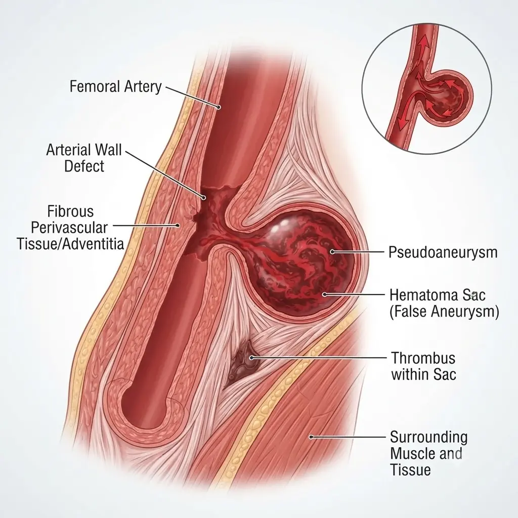
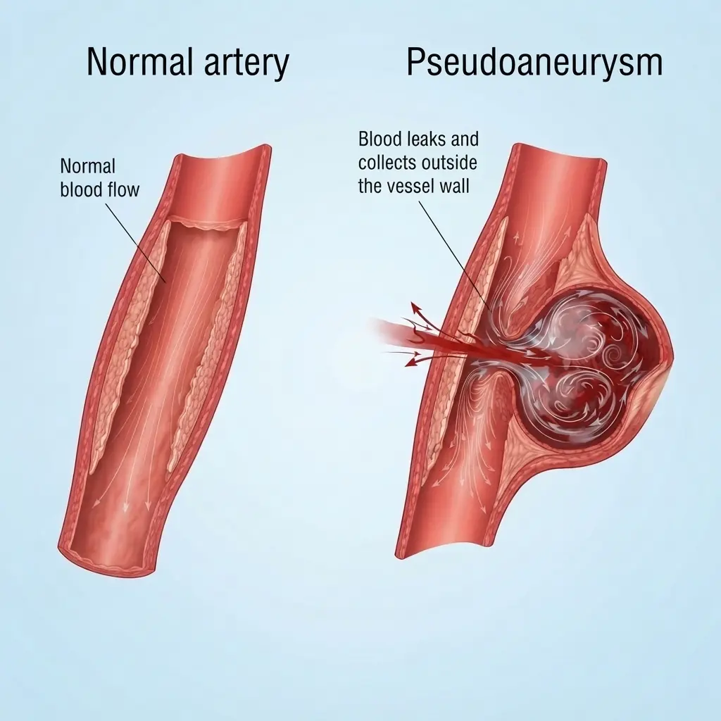
A peripheral pseudoaneurysm develops when blood leaks through an injured or weakened blood vessel wall and collects in the surrounding tissue while remaining connected to the artery. It may occur after a vascular procedure, an injury, or other damage to a blood vessel.

Dr. Anadi Gupta, MD, FVIR, EBIR

Consultant, Sir Ganga Ram Hospital

Peripheral Pseudoaneurysm Embolization
Peripheral Pseudoaneurysms

Understanding Peripheral Pseudoaneurysms

What is a peripheral pseudoaneurysm?

A pseudoaneurysm, sometimes called a false aneurysm, occurs when blood escapes through a defect in an artery wall and forms a contained collection outside the normal vessel layers. The collection remains connected to the artery, allowing blood to flow into it with each heartbeat.Peripheral pseudoaneurysms can develop in arteries supplying the arms, legs, or other parts of the body. They may occur after catheter-based procedures, vascular surgery, accidental injury, or trauma. Some develop near a previous arterial access site.

Common Problems

What is pseudoaneurysm embolization?

Image-Guided Treatment to Control Abnormal Blood Flow

Peripheral pseudoaneurysm embolization is a minimally invasive procedure used to block blood flow into a pseudoaneurysm by targeting the responsible vessel or vessels. The objective is to prevent further filling of the collection while preserving circulation to the surrounding tissues whenever possible.
Under imaging guidance, an interventional specialist identifies the pseudoaneurysm and its connection to the artery. A catheter may be introduced through a blood vessel to deliver an appropriate embolic material, such as coils or another agent, depending on the anatomy and clinical situation.
Signs and Symptoms of a Pseudoaneurysm

Who may benefit from treatment?

When Is Pseudoaneurysm Treatment Recommended?

A vascular specialist may recommend further evaluation or treatment for patients with:
  • A pseudoaneurysm that is enlarging or causing persistent pain.
  • A pseudoaneurysm that develops after an arterial catheter-based procedure.
  • A pseudoaneurysm following an injury or vascular intervention.
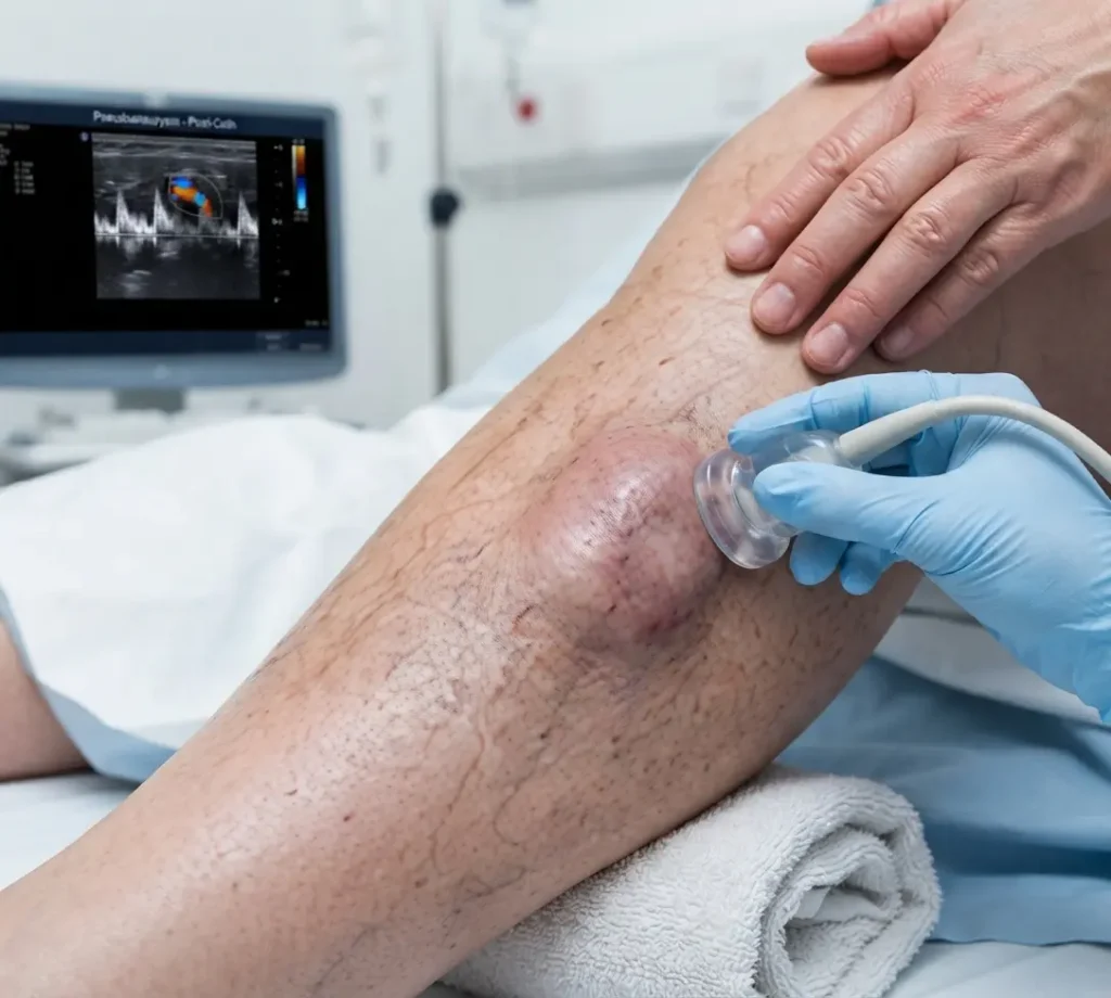
  • Continued blood flow into the pseudoaneurysm on Doppler ultrasound.
  • Pressure on nearby nerves or other structures.
  • A pseudoaneurysm associated with impaired circulation or other complications.
  • May stop or reduce abnormal blood flow into the pseudoaneurysm.
  • Uses image guidance to target the responsible blood vessels.
  • May avoid open surgery in selected cases.
  • May help reduce the risk of further enlargement or bleeding when successful treatment is achieved.
  • Can be tailored to the pseudoaneurysm’s location and the surrounding arterial anatomy.
  • Bleeding, bruising, or discomfort at the catheter access site.
  • Injury to the treated blood vessel.
  • Incomplete closure or recurrence of the pseudoaneurysm.
  • Unintended blockage of a nearby artery or reduced blood flow to surrounding tissues.
  • Blood clot formation or circulation-related complications.
  • Infection, depending on the clinical circumstances.
     

How is the procedure performed?

The Peripheral Pseudoaneurysm Embolization Process

Consultation and Assessment

Dr. Anadi Gupta reviews your symptoms, medical history, recent procedures or injuries, medication use, and relevant imaging.

Diagnostic Imaging

Doppler ultrasound is commonly used to confirm the pseudoaneurysm, assess its size, and identify the connection with the artery. CT angiography or catheter angiography may be required in selected cases.

Personalized Treatment Planning

The specialist evaluates the location of the pseudoaneurysm and the circulation to surrounding tissues. Treatment options and their potential risks are discussed.

Image-Guided Intervention

If embolization is appropriate, a catheter may be guided to the target vessels under imaging guidance. Coils or other suitable embolic materials are used to stop blood flow into the pseudoaneurysm. The exact technique depends on the anatomy and the need to preserve the main artery.

Confirmation of Treatment

Imaging is used to assess whether blood flow into the pseudoaneurysm has been controlled and whether circulation to the surrounding tissues remains adequate.

Recovery and Follow-Up

The access site and treated area are monitored after the procedure. Follow-up examination or Doppler ultrasound may be recommended to confirm continued closure and identify any recurrence.

1

Why consult Dr. Anadi Gupta?

Specialized Vascular & Endovascular Care

Dr. Anadi Gupta, MD, FVIR, EBIR, is a Consultant at Sir Ganga Ram Hospital with a focus on vascular and endovascular interventions.His areas of practice include peripheral pseudoaneurysm embolization, vascular malformation embolization and sclerotherapy, peripheral arterial disease angioplasty and stenting, DVT thrombectomy and catheter-directed thrombolysis, central venous angioplasty and stenting, and dialysis access interventions.

ADVANCED VASCULAR & ENDOVASCULAR CARE

Need Evaluation for a Peripheral Pseudoaneurysm?

Consult Dr. Anadi Gupta at Sir Ganga Ram Hospital to understand your diagnosis and explore suitable treatment options, including image-guided embolization when appropriate.

Frequently Asked Questions

Peripheral Pseudoaneurysm Embolization

What is a peripheral pseudoaneurysm?

A peripheral pseudoaneurysm occurs when blood escapes through a defect in an artery wall and collects in surrounding tissue while remaining connected to the artery. It may develop after an injury or vascular procedure.

Some pseudoaneurysms remain stable, while others can enlarge, bleed, compress nearby structures, or affect circulation. Prompt assessment helps determine the level of risk and the need for treatment.

Doppler ultrasound is commonly used to identify the pseudoaneurysm and assess blood flow. CT angiography or catheter angiography may be required in more complex cases.

No. Treatment may include observation, ultrasound-guided compression, thrombin injection, embolization, covered stent placement, or surgery. The choice depends on the pseudoaneurysm's characteristics and the patient's condition.

Embolization is generally a minimally invasive, image-guided procedure performed through a small catheter access point. The exact approach and anesthesia depend on the individual case.

Recurrence or incomplete closure can occur. Follow-up imaging may be recommended to confirm that blood flow into the pseudoaneurysm has stopped.

Urgency depends on the pseudoaneurysm's size, location, symptoms, and risk of complications. Rapid enlargement, severe pain, uncontrolled bleeding, fainting, or a cold or numb limb requires emergency medical assessment.